7.3 Continued
In cotransport, an ATP pump that transports a specific solute indirectly drives the active transport of other substances. In this process, the substance that was initially pumped across the membrane will do work, providing energy for the transport of another substance against its concentration gradient, as it leaks back across the membrane w/its concentration gradient.
Large molecules are moved across the cell membrane through exocytosis and endocytosis. In exocytosis, vesicles from the cell's interior fuse w/the cell membrane expelling their contents to the exterior. In endocytosis, the cell forms new vesicles from the plasma membrane; this is basically the reverse of exocytosis, and this process allows the cell to take in macromolecules.
There are three types of endocytosis:
1. Phagocytosis (remember: phag = to eat) occurs when the cell wraps pseudopodia around the substance and packages it within a large vesicle formed by the membrane.
2. In pinocytosis (remember: pino = to drink), the cell takes in small droplets of extracellular fluid in small vesicles. Pinocytosis is not specific – no target molecules are taken in, in this process.
3. Receptor-mediated endocytosis is a very specific process. Certain substances (ligands) bind to specific receptors on the cell's surface (these receptors are usually clustered in coated pits), and this causes a vesicle to for around the substance and then to pinch off into the cytoplasm.
CHAPTER SEVEN ESSAY TOMORROW!!!!!!!
For review, here is a brief outline of cellular transportation:
I. Passive Transport
A. Diffusion
i. High to low concentration
ii. MOST molecules that can diffuse are nonpolar and hydrophobic
B. Facilitated Diffusion
i. Transport proteins
a. Channel proteins
b. Carrier proteins
ii. High to low
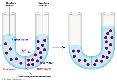
C. Osmosis
i. Water
ii. Selectively permeable membrane
iii. Hypotonic to hypertonic
II. Active Transport
A. ATP required
i. Low to high
B. Primary Active Transport
i. Sodium-potassium pump (3:2 ratio of Na to K)
C. Cotransport
III. Other
A. Exocytosis
B. Endocytosis
i. Phagocytosis
ii. Pinocytosis
iii. Receptor-mediated
a. SPECIFIC
GOOD LUCK ON THAT ESSAY!!!!
Chapter 36
Resource Acquisition and Transport in Vascular Plants
Section 1
An Overview of Transport Mechanisms in Plants:
Three kinds of transport occur in plants:
1. The uptake and loss of water and minerals from individual cells, for instance in a root cell or leaf cell.
2. Transport of substances short distances, from cell to cell
3. Transport of sap within the xylem and phloem, throughout the entire plant
In plants, the uptake of water across cell membranes occurs through osmosis, the passive transport of water across a membrane.
The water potential is defined as the combined effects of solute concentration and the pressure that the cell wall contributes.
Turgor pressure is the pressure exerted against the cell wall when the cell is filled w/water.
Aquaporins are the channels in the plant cell walls specifically designed for the passage of water. How might this affect osmosis?
Plants have a tonoplast surrounding their vacuoles; the tonoplast regulates molecules going into and out of the vacuole.
The symplast is a continuation of cytoplasm that is connected by plasmodesmata between cells.
The apoplast is the nonliving continuum that is formed by the extracellular pathway formed by the continuous matrix of cell walls.
DONT FORGET: CHAPTER 36 VOCAB DUE TOMORROW!!!!


