Hey girls, we had some random things to review in class today including signal transduction so hopefully this clears up any confusion in that area.
External signals are converted into responses within the cell
Evolution of Cell Signaling:
Signaling in microbes has much in common with processes in multicellular organisms, suggesting an early origin.
Local and Long–Distance Signaling:
In local signaling, animal cells may communicate by direct contact or by secreting local regulators, such as growth factors or neurotransmitters. For signaling over long distances, both animals and plants use hormones; animals also signal along nerve cells.
The Three Stages of Cell Signaling:
Earl Sutherland discovered how the hormone epinephrine acts on cells. The signal molecule epinephrine binds to receptors on a cell′s surface (reception), leading to a series of changes in the receptor and other molecules inside the cell (transduction) and finally to the activation of an enzyme that breaks down glycogen (response).
Reception: A signal molecule binds to a receptor protein, causing it to change shape
The binding between signal molecule (ligand) and receptor is highly specific. A conformational change in a receptor is often the initial transduction of the signal
Intracellular Receptors:
Intracellular receptors are cytoplasmic or nuclear proteins. Signal molecules that are small or hydrophobic and can readily cross the plasma membrane use these receptors.
Receptors in the Plasma Membrane:
A G–protein–linked receptor is a membrane receptor that works with the help of a cytoplasmic G protein. Ligand binding activates the receptor, which then activates a specific G protein, which activates yet another protein, thus propagating the signal along a signal transduction pathway.
Receptor tyrosine kinases react to the binding of signal molecules by forming dimers and then adding phosphate groups to tyrosines on the cytoplasmic side of the other subunit of the receptor. Relay proteins in the cell can then be activated by binding to different phosphorylated tyrosines, allowing this receptor to trigger several pathways at once.
Specific signal molecules cause ligand–gated ion channels in a membrane to open or close, regulating the flow of specific ions.
Transduction: Cascades of molecular interactions relay signals from receptors to target molecules in the cell
Signal Transduction Pathways:
At each step in a pathway, the signal is transduced into a different form, commonly a conformational change in a protein.
Protein Phosphorylation and Dephosphorylation:
Many signal transduction pathways include phosphorylation cascades, in which a series of protein kinases each add a phosphate group to the next one in line, activating it. Phosphatase enzymes soon remove the phosphates.
Small Molecules and Ions as Second Messengers:
Second messengers, such as cyclic AMP (cAMP) and Ca2+, diffuse readily through the cytosol and thus help broadcast signals quickly. Many G proteins activate adenylyl cyclase, which makes cAMP from ATP. Cells use Ca2+ as a second messenger in both G–protein and tyrosine kinase pathways. The tyrosine kinase pathways can also involve two other second messengers, DAG and IP3. IP3 can trigger a subsequent increase in Ca2+ levels.
Response: Cell signaling leads to regulation of cytoplasmic activities or transcription
Cytoplasmic and Nuclear Responses:
In the cytoplasm, signaling pathways regulate, for example, enzyme activity and cytoskeleton rearrangement. Other pathways regulate genes by activating transcription factors, proteins that turn specific genes on or off.
Fine–Tuning of the Response:
Each catalytic protein in a signaling pathway amplifies the signal by activating multiple copies of the next component of the pathway; for long pathways, the total amplification may be a millionfold or more. The particular combination of proteins in a cell gives the cell great specificity in both the signals it detects and the responses it carries out. Scaffolding proteins can increase signal transduction efficiency. Pathway branching and cross–talk further help the cell coordinate incoming signals. Signal response is terminated quickly by the reversal of ligand binding.
Quizlet:
1. Amplification of a chemical signal occurs when
a. a receptor in the plasma membrane activates several G– protein molecules while a signal molecule is bound to it.
b. a cAMP molecule activates one protein kinase molecule before being converted to AMP.
c. phosphorylase and phosphatase activities are balanced.
d. receptor tyrosine kinases dimerize upon ligand binding.
e. both a and d occur.
2. Which of the following provides the best evidence that cell–signaling pathways evolved early in the history of life?
a. They are seen in “primitive” cells such as yeast.
b. Yeast cells signal each other for mating.
c. Signal transduction molecules found in distantly related organisms are similar.
d. Signals can be sent long distances by cells.
e. Most signals are received by cell surface receptors.
3. Consider this pathway: epinephrine → G–protein–linked receptor → G protein → adenylyl cyclase → cAMP. Identify the second messenger.
a. cAMP
b. G protein
c. GTP
d. adenylyl cyclase
e. G–protein–linked receptor
4. Which observation suggested to Sutherland the involvement of a second messenger in epinephrine′s effect on liver cells?
a. Enzymatic activity was proportional to the amount of calcium added to a cell–free extract.
b. Receptor studies indicated that epinephrine was a ligand.
c. Glycogen breakdown was observed only when epinephrine was administered to intact cells.
d. Glycogen breakdown was observed when epinephrine and glycogen phosphorylase were combined.
e. Epinephrine was known to have different effects on different types of cells.
5. Binding of a signal molecule to which type of receptor leads directly to a change in the distribution of anions and/or cations on opposite sides of the membrane?
a. receptor tyrosine kinase
b. G–protein–linked receptor
c. phosphorylated receptor tyrosine kinase dimer
d. ligand–gated ion channel
e. intracellular receptor
6. Protein phosphorylation is commonly involved with all of the following except
a. regulation of transcription by extracellular signal molecules.
b. enzyme activation.
c. activation of G–protein–linked receptors.
d. activation of receptor tyrosine kinases.
e. activation of protein kinase molecules.
7. Signal transduction pathways benefit cells for all of the following reasons except
a. they help cells respond to signal molecules that are too large or too polar to cross the plasma membrane.
b. they enable different cells to respond appropriately to the same signal.
c. they help cells use up phosphate generated by ATP breakdown.
d. they can amplify a signal.
e. variations in the signal transduction pathways can enhance response specificity.
8. Phosphorylation cascades involving a series of protein kinases are useful for cellular signal transduction because
a. they are species specific.
b. they always lead to the same cellular response.
c. they amplify the original signal manyfold.
d. they counter the harmful effects of phosphatases.
e. the number of molecules used is small and fixed.
9. The activation of receptor tyrosine kinases is always characterized by
a. dimerization and phosphorylation.
b. IP3 binding.
c. a phosphorylation cascade.
d. GTP hydrolysis.
e. channel protein conformational change.
10. Lipid–soluble signal molecules, such as testosterone, cross the membranes of all cells but affect only target cells because
a. only target cells retain the appropriate DNA segments.
b. intracellular receptors are present only in target cells.
c. most cells lack the Y chromosome required.
d. only target cells possess the cytosolic enzymes that transduce the testosterone.
e. only in target cells is testosterone able to initiate the phosphorylation cascade leading to activated transcription factor.
Answers:
1. A
2. C
3. A
4. C
5. D
6. C
7. C
8. C
9. A
10. B
Tuesday, May 5, 2009
Saturday, May 2, 2009
Review: Protein Synthesis!
I thought that a review on protein synthesis, including transcription, RNA processing and translation would be helpful. On Friday B block didn't really have class, and in C block we just watched a movie. The chapter 50 packet is now due on Monday, and keep reviewing!
PROTEIN SYNTHESIS
Protein synthesis is the process that describes how enzymes and other proteins are made from DNA.
The three steps in protein synthesis are transcription, RNA processing, and translation.
Transcription:
This is the process by which DNA makes RNA. The three types of RNA are mRNA, tRNA, and rRNA. Messenger RNA (mRNA) carries messages directly from DNA to the cytoplasm and varies in length, depending on the length of the message. Transfer RNA (tRNA) is shaped like a cloverleaf and carries amino acids to the mRNA at the ribosome. Ribosomal RNA (rRNA) is strcutural, this makes up the ribosome, which is formed in the nucleolus.
Transcription consists of three stages: initiaition, elongation, and termination.
At the start of initiation an enzyme, RNA polymerase, recognizes and binds to DNA at the promoter region. This begins to unzip the DNA into two strands. A promoter region for mRNA transcriptions often contains the sequence T-A-T-A (the TATA box). The completed assembly of transcription factors and RNA polymerase bound to the promoter is called the transcription initiation complex. Once RNA polmerase is attached to promoter, DNA transcription of the DNA template begins.
Elongation occurs as the RNA polymerase unzips the DNA and assembles RNA nucleotides using one strand of the DNA as a template. As in DNA replication , elongation of the RNA molecule occurs in the 5' --> 3' direction. In contrast to DNA replication, new nucleotides (rather than DNA nucleotides), and only on DNA strand in transcribed.
Termination is the final stage. It continues for a short distance after the RNA polymerase transcribes the termination sequence. At this point, mRNA is cut free from the DNA template. In eukaryotes, the termination region often ocntains the DNA sequence AAAAAAA.
 RNA Processing
Before the newly formed RNA strand is shipped out to the ribosome in the cytoplasm, it is altered or processed by a series of enzymse.
A 5' cap consisting of a modified guanine nucleotide is added to the 5' end. This cap helps protect the RNA strand from degradation by hydrolytic enzymes and also helps the RNA strands bind to the ribosome in the cytolpasm.
A poly-A tail is attahced to the 3' end of the mRNA. The tail consists of 30 to 200 adenine nucleotides. The tail protects the RNA strand from degradation by hydrolytic enzymes, helps the ribosome attach to the RNA, and facilitates the release of the RNA into the cytplasm.
Noncoding regions of the mRNA called introns or intervening sequences are removed by the snRNPs, small nuclear ribonucleoproteins, and splicesomes.This removal allows only exons, which are expressed sequences, to leaves the nucleus. As a result of this processing, the mRNA that leaves the nucleus is a great deal shorter than the original transcription unit.

Translation
After transcription, the mRNA, tRNA, and ribosomal subunits are transported across the nuclear envelope and into the cytoplasm. In the cytoplasm, amino acids attach to the 3' end of the tRNA's, forming an aminoacyl-tRNA. The reaction requires an enzyme specific to each tRNA and the energy from one ATP. The amino acid-tRNA bond that results is a high-energy bond, creating a activated amino acid.
Like transcription, translation is categorized into three steps: intiation. elongation, and termination. Energy for translation is provided by several GTP molecules. GTP acts as an energ supplier in the same manner as ATP.
Intiation begins with when mRNA becomes attached to a subunit of the ribosome. The first codon is always AUG. It codes for methione and must be positioned correctly in order for transcription of an amino acid sequence to begin. At this point, the ribosomal subunit is a complex of mRNA, AUG, the tRNA anticodon, UAC, the amino acid methione.
Elongation continues as tRNA brings amino acids to the ribosome and a polypeptide chain is formed.
Termination of an mRNA strand is complete when a ribosome reaches one of three termination or stop codons. A release factor hydrolyzes the bond between ther tRNA and the last amino acid of the polypeptide chain. The polypeptide is freed from the ribosome, and the mRNA is broken down.
 Quiz:
Quiz:
1) If AUU is the codon, what is the anticodon?
a. AUU
b. TAA
c. UUA
d. UAA
2) What are the regions of DNA called that code for proteins?
a. introns
b. codons
c. anticodons
d. exons
3) The end products of translation are
a. polypeptides
b. amino acids
c. RNA
d. DNA
4) Which if the following contains a code for a protein?
a. mRNA
b. RNA polymerase
c. rRNA
d. tRNA
5) Proteins synthesis consists of all the following steps except:
a. translation
b. transcription
c. replication
d. elongation
ANSWERS: 1)D 2)D 3)A 4)A 5)C
Just keep reviewing the rest of the week!!!! :-)
PROTEIN SYNTHESIS
Protein synthesis is the process that describes how enzymes and other proteins are made from DNA.
The three steps in protein synthesis are transcription, RNA processing, and translation.
Transcription:
This is the process by which DNA makes RNA. The three types of RNA are mRNA, tRNA, and rRNA. Messenger RNA (mRNA) carries messages directly from DNA to the cytoplasm and varies in length, depending on the length of the message. Transfer RNA (tRNA) is shaped like a cloverleaf and carries amino acids to the mRNA at the ribosome. Ribosomal RNA (rRNA) is strcutural, this makes up the ribosome, which is formed in the nucleolus.
Transcription consists of three stages: initiaition, elongation, and termination.
At the start of initiation an enzyme, RNA polymerase, recognizes and binds to DNA at the promoter region. This begins to unzip the DNA into two strands. A promoter region for mRNA transcriptions often contains the sequence T-A-T-A (the TATA box). The completed assembly of transcription factors and RNA polymerase bound to the promoter is called the transcription initiation complex. Once RNA polmerase is attached to promoter, DNA transcription of the DNA template begins.
Elongation occurs as the RNA polymerase unzips the DNA and assembles RNA nucleotides using one strand of the DNA as a template. As in DNA replication , elongation of the RNA molecule occurs in the 5' --> 3' direction. In contrast to DNA replication, new nucleotides (rather than DNA nucleotides), and only on DNA strand in transcribed.
Termination is the final stage. It continues for a short distance after the RNA polymerase transcribes the termination sequence. At this point, mRNA is cut free from the DNA template. In eukaryotes, the termination region often ocntains the DNA sequence AAAAAAA.
Before the newly formed RNA strand is shipped out to the ribosome in the cytoplasm, it is altered or processed by a series of enzymse.
A 5' cap consisting of a modified guanine nucleotide is added to the 5' end. This cap helps protect the RNA strand from degradation by hydrolytic enzymes and also helps the RNA strands bind to the ribosome in the cytolpasm.
A poly-A tail is attahced to the 3' end of the mRNA. The tail consists of 30 to 200 adenine nucleotides. The tail protects the RNA strand from degradation by hydrolytic enzymes, helps the ribosome attach to the RNA, and facilitates the release of the RNA into the cytplasm.
Noncoding regions of the mRNA called introns or intervening sequences are removed by the snRNPs, small nuclear ribonucleoproteins, and splicesomes.This removal allows only exons, which are expressed sequences, to leaves the nucleus. As a result of this processing, the mRNA that leaves the nucleus is a great deal shorter than the original transcription unit.

Translation
After transcription, the mRNA, tRNA, and ribosomal subunits are transported across the nuclear envelope and into the cytoplasm. In the cytoplasm, amino acids attach to the 3' end of the tRNA's, forming an aminoacyl-tRNA. The reaction requires an enzyme specific to each tRNA and the energy from one ATP. The amino acid-tRNA bond that results is a high-energy bond, creating a activated amino acid.
Like transcription, translation is categorized into three steps: intiation. elongation, and termination. Energy for translation is provided by several GTP molecules. GTP acts as an energ supplier in the same manner as ATP.
Intiation begins with when mRNA becomes attached to a subunit of the ribosome. The first codon is always AUG. It codes for methione and must be positioned correctly in order for transcription of an amino acid sequence to begin. At this point, the ribosomal subunit is a complex of mRNA, AUG, the tRNA anticodon, UAC, the amino acid methione.
Elongation continues as tRNA brings amino acids to the ribosome and a polypeptide chain is formed.
Termination of an mRNA strand is complete when a ribosome reaches one of three termination or stop codons. A release factor hydrolyzes the bond between ther tRNA and the last amino acid of the polypeptide chain. The polypeptide is freed from the ribosome, and the mRNA is broken down.
1) If AUU is the codon, what is the anticodon?
a. AUU
b. TAA
c. UUA
d. UAA
2) What are the regions of DNA called that code for proteins?
a. introns
b. codons
c. anticodons
d. exons
3) The end products of translation are
a. polypeptides
b. amino acids
c. RNA
d. DNA
4) Which if the following contains a code for a protein?
a. mRNA
b. RNA polymerase
c. rRNA
d. tRNA
5) Proteins synthesis consists of all the following steps except:
a. translation
b. transcription
c. replication
d. elongation
ANSWERS: 1)D 2)D 3)A 4)A 5)C
Just keep reviewing the rest of the week!!!! :-)
Tuesday, April 28, 2009
Chapter 43- The Body's Defenses
In innate immunity, recognition and response rely on shared pathogen traits
Innate immune responses include barrier defenses as well as defenses to combat pathogens that end the body
1. Barrier defenses skin and the mucous membranes cover the surface and line the openings of the animal body, and they provide an external barrier against infecting agents
First Line of Defense
Specifically, skin, antimicrobial proteins are in the saliva, cilia lines the lungs to sweep invaders out, gastric juice of the stomach kills most microbes, and symbiotic bacteria in the digestive tract also serve as first line of denfense
Microbes that get through the skin-- for instance, in a cut-- encounter certain types of white blood cells called neutrophils that ingest and destroy them in a process called phagocytosis
Here is a link to a video showing neutrophil phagocytosis
http://esgweb1.nts.jhu.edu/cellbio/devreotes/neutrophil.mov
2. Cellular innate defenses combat pathogens that get through the skin-- for example, in a cut. They include phagocytic white blood cells and antimicrobial proteins.
Phagocytic White Blood Cells
Antimicrobial Proteins
Inteferon proteins provide innate defense against viral infections. They cause cells adjacent to injected cells to produce substances to inhibit viral replication
The complement system consists of roughly 30 proteins with a variety of functions. One function is to lyse invading cells.
The major histocompatibility complex, or MHC, is the mechanism by which the immune system is able to differentiate between self and nonself cells. The MHC is a collection of glycoproteins that exists on the membranes of all body cells.
3. The inflammatory response occurs where physical injury occurs to a tissue, and occurs in response to chemical signals (Second line of defense). For example, histamines are released by basophils and most cells (two ypes of leukocytes) in response to injury. Histamines trigger the dilation and permeability of nearby capillaries. This aids in delivering clotting agents (white blood cells) to the injured area. Vasodilation causes redness, and increase in temperature, and and swelling. Histamines cause watery, itchy eyes, and sneezing. We fight histamine reactions with antihistamines.

The third line of defense is the immune response and it differs from the inflammatory response in that it targets specific antigens.
4. Natural Killer (NK) cells help recognize and remove diseased cells
In acquired immunity, lymphocyte receptors provide pathogen- specific recognition
Vertebrates have two types of: B lymphocytes (B cells) which proliferate in the bone marrow, and T lymphocytes (T cells) where lymphocytes mature in the thymus. They circulate through the blood and lymph, and both recognize particular microbes and are said to show specificity.
Antigens are foreign molecules that elicit a response by lymphocytes
Antibodies are proteins secreted by B cells in an immune response. Each anitibody is specific to a particular antigen. There are five classes of anitbodies (or immunoglobins): IgA, IgD, IgE, IgG, IgM.
Antigen receptors are located on the antigen and allow B and T cells to recognize them as antigen receptors on T cells are called T cell receptors, which combat the antigen, and the other consists of memory cells, which are long-lived and bear receptors for the same antigen.
Plasma cells are B cells that release their specific antibodies which then circulate through the body, binding to antigens.
Memory cells are lon-lived B cells that do not release their antibodies in response to the immediate antigen invasion. Instead, the memory cells circulate in the body and respond quickly to eliminate any subsequent invasion by the same antigen. This mechanism provides immunity to many diseases after the first occurence of the disease.
Here is a link to a video about white blood cells, T-cells, NK cells, and cancer cells
http://www.metacafe.com/watch/yt-aSpRpf1Ny9A/immune_system_white_blood_cells_t_cells_cancer_cells/
Multiple Choice Questions
1. Which if these cells produce and secrete antibodies?
a) macrophages
b) cytotoxic T cells
c) helper T cells
d) bacterial cells
e) plasma cells
2. What kind of white blood cells defend against parasitic invaders?
a) basophils
b) monocytes
c) eosinophils
d) neutrophils
e) histamines
3. ________ proliferate in the bone marrow and ________ mature in the thymus.
a) B cells, T cells
b) NK cells, T cells
c) T cells, B cells
d) B cells, NK cells
e) NK cells, B cells
4. Histamine is secreted by ________.
a) eosinophils
b) monocytes
c) basophils
d) T lymphocytes
e) B lymphocytes
Answers: 1. e, 2. c, 3. a, 4. c
Notes reposted from March 30th, due to technical difficulties with my blog account.
In innate immunity, recognition and response rely on shared pathogen traits
Innate immune responses include barrier defenses as well as defenses to combat pathogens that end the body
1. Barrier defenses skin and the mucous membranes cover the surface and line the openings of the animal body, and they provide an external barrier against infecting agents
First Line of Defense
Specifically, skin, antimicrobial proteins are in the saliva, cilia lines the lungs to sweep invaders out, gastric juice of the stomach kills most microbes, and symbiotic bacteria in the digestive tract also serve as first line of denfense
Microbes that get through the skin-- for instance, in a cut-- encounter certain types of white blood cells called neutrophils that ingest and destroy them in a process called phagocytosis
Here is a link to a video showing neutrophil phagocytosis
http://esgweb1.nts.jhu.edu/cellbio/devreotes/neutrophil.mov
2. Cellular innate defenses combat pathogens that get through the skin-- for example, in a cut. They include phagocytic white blood cells and antimicrobial proteins.
Phagocytic White
Blood Cells- Neutrophils are white blood cells that ingest and destroy microbes in a process called phagocytosis
- Monocytes are another type of phagocytic leukocyte. They migrate into tissues and develop into macrophages, which are giant phagocytic cells
- Eosinophils are leukocytes that defend against parasitic invaders such as worms by positioning themsleves near the parasite's wall and discharging hydrolytic enzymes
Antimicrobial Proteins
Inteferon proteins provide innate defense against viral infections. They cause cells adjacent to injected cells to produce substances to inhibit viral replication
The complement system consists of roughly 30 proteins with a variety of functions. One function is to lyse invading cells.
The major histocompatibility complex, or MHC, is the mechanism by which the immune system is able to differentiate between self and nonself cells. The MHC is a collection of glycoproteins that exists on the membranes of all body cells.
3. The inflammatory response occurs where physical injury occurs to a tissue, and occurs in response to chemical signals (Second line of defense). For example, histamines are released by basophils and most cells (two ypes of leukocytes) in response to injury. Histamines trigger the dilation and permeability of nearby capillaries. This aids in delivering clotting agents (white blood cells) to the injured area. Vasodilation causes redness, and increase in temperature, and and swelling. Histamines cause watery, itchy eyes, and sneezing. We fight histamine reactions with antihistamines.
The third line of defense is the immune response and it differs from the inflammatory response in that it targets specific antigens.
4. Natural Killer (NK) cells help recognize and remove diseased cells
In acquired immunity, lymphocyte receptors provide pathogen- specific recognition
Vertebrates have two types of: B lymphocytes (B cells) which proliferate in the bone marrow, and T lymphocytes (T cells) where lymphocytes mature in the thymus. They circulate through the blood and lymph, and both recognize particular microbes and are said to show specificity.
Antigens are foreign molecules that elicit a response by lymphocytes
Antibodies are proteins secreted by B cells in an immune response. Each anitibody is specific to a particular antigen. There are five classes of anitbodies (or immunoglobins): IgA, IgD, IgE, IgG, IgM.
Antigen receptors are located on the antigen and allow B and T cells to recognize them as antigen receptors on T cells are called T cell receptors, which combat the antigen, and the other consists of memory cells, which are long-lived and bear receptors for the same antigen.
Plasma cells are B cells that release their specific antibodies which then circulate through the body, binding to antigens.
Memory cells are lon-lived B cells that do not release their antibodies in response to the immediate antigen invasion. Instead, the memory cells circulate in the body and respond quickly to eliminate any subsequent invasion by the same antigen. This mechanism provides immunity to many diseases after the first occurence of the disease.
Here is a link to a video about white blood cells, T-cells, NK cells, and cancer cells
http://www.metacafe.com/watch/yt-aSpRpf1Ny9A/immune_system_white_blood_cells_t_cells_cancer_cells/
Multiple Choice Questions
1. Which if these cells produce and secrete antibodies?
a) macrophages
b) cytotoxic T cells
c) helper T cells
d) bacterial cells
e) plasma cells
2. What kind of white blood cells defend against parasitic invaders?
a) basophils
b) monocytes
c) eosinophils
d) neutrophils
e) histamines
3. ________ proliferate in the bone marrow and ________ mature in the thymus.
a) B cells, T cells
b) NK cells, T cells
c) T cells, B cells
d) B cells, NK cells
e) NK cells, B cells
4. Histamine is secreted by ________.
a) eosinophils
b) monocytes
c) basophils
d) T lymphocytes
e) B lymphocytes
Answers: 1. e, 2. c, 3. a, 4. c
Notes reposted from March 30th, due to technical difficulties with my blog account.
Monday, April 27, 2009
Review: Cellular Respiration
Today in class we did interactive slides on the smart board to start our review for the AP test.


Here are the topics the slides covered:
-sources and sinks
-hydrogen bonds
-the four properties of water:
+cohesion and adhesion (transpiration)
+the fact that water is less dense as a solid than as a liquid
+surface tension
+high speific heat
-modes of selection
+stabilizing
+directoinal
+disruptive
-speciation (*a lot of people seemed to need to review this one)
-macromolecules
-apoplastic and symplastic routes
-recombinant DNA and vectors
-guttation, xenophytes, C3 C4 and CAM plants
-stages of mitosis
-guttation, xenophytes, C3 C4 and CAM plants
-stages of mitosis
-animal junctions
+desmosome
+tight junction
+gap junction
-plant junctions
+plasmodesmata
-monomers, polymers, and bonds
-bacteriophages
-the structure of a protein
I, personally, feel that I need to brush up on CELLULAR RESPIRATION. Here is a quick review. Hopefully it helps everyone out!
Cellular respiration is the breakdown of sugars that occurs in teh presence of oxygen. Carbohydrates, fats, and proteins can all be broken down to release energy in cellular repsiration. However, glucose is the primary nutrient fuel molecule that is used in cellular respiration to release energy.
Below is the standard equation for cellular respiration:
Cellular respiration is aerobic respiration (oxygen is present).
Glycolysis is the first process in cellular respiration:
Glycolysis is the decomposition of glucose to two pyruvate molecules. The 6-carbon glucose molecule is split into two 3-carbon sugars through a long series of steps.
There is an ATP consuming phase and an ATP producing phase.
First, 2 ATP are consumed. In the next phase, 4 ATP are produced as well as 2 NADH. The net gain of ATP is 2.
*NADG is a conenzyme that forms when NAD+ combines with two energy-rich electrons and H+.
The Krebs Cycle is the second process:
It occurs in the mitochondrial matrix. In it, glucose is completely broken down and the final product is CO2.
The cycle has 8 steps, and each is catalyzed by a different enzyme:
1. Acetyl CoA adds iots two-carbon acetyl group to oxoaloacetate, producing citrate.
2. Citrate is converted to its isomer, isocitrate, by removal of one water molecule and addition of another.
3. Isocitrate is oxidized, reducing NAD+ to NADH. Then the resultinf compound loses a CO2 molecule.
4. Another CO2 is lostm and the resulting compound is oxidized, reducing NAD+ to NADH. THe remaining molecule is then attached to coenzyme A by an unstable bond.
5. CoA is displaced by a phosphate group, which is transferred to GDP, forming GTP, a molecule with functions similar to ATP.
6. Two hydrogens are transferred to FAD, forming FADH2 and oxidizing succinate.
7. Addition of a water molecule rearranges bonds in the substrate.
8. The substrate is oxidized, reducing NAD+ to NADH and regenerating oxaloacetate.
Each cycling requires the input of a 2-carbon acetyl co-a molecule, and two carbons are released in the course of the cyle as CO2.
For every 1 turn of the cycle, the net results are:
2 CO2
1 ATP
3 NADH
1 FADH2
For every 2 turns of the cylce, the net results are:
4 CO2
4 CO2
2 ATP
6 NADH
2 FADH2
 **Both glycolysis and the Krebs Cycle are substrate level phosphorylation. This means that a phosphate group and its associated energy is transferred to ADP to from ATP. The substrate molecule (the one with the phosphate group) donates the high energy phosphate group.
**Both glycolysis and the Krebs Cycle are substrate level phosphorylation. This means that a phosphate group and its associated energy is transferred to ADP to from ATP. The substrate molecule (the one with the phosphate group) donates the high energy phosphate group.Oxidative phosphorylation is the last process of the cellular respiration:
Oxidative phosphorylation is the process of extracting ATP from NADH and FADH2. Electrons from NADH and FADH2 pass along an electron transport chain.
The electron transport chain consists of molecules (mainly proteins) embedded in teh inner mitochondrial membrane. Sitting on top of these proteins embedded in the membrane are associated molecules that are alternately reduced and oxidized as the accept and donate electrons.
* In oxidation, electrons are loast. In reduction, they are added.
The first electron acceptor in the ETC is a flavoprotein called flavin mononucleotide (FMN) and it accepts and electron from NADH.
The electron is passd down a series of molecules to oxygen, the final electron acceptor. Then, it is combined with hydrogen atoms to form water.
*FADH2 and NADH both donate electrons in the chain
The ETC does not make any ATP itself. Its reactions are coupled to others to make ATP in chemiosmosis.
In chemiosmosis, a hydrogen pump pumps H+ ions against their concentration gradient across the mitochondrial membrane. This creates a proton gradient. Protein complexes called ATP synthases embedded in the membrane use the energy from the proton gradient. The H+ ions flow back into the mitochondrial matrix through the ATP synthase. The flow drives the oxidative phosphorylation of ADP to ATP. Oxidative phosphorylation results in about 34 ATP molecules.
This makes the total number of ATP produced by cellular respiration about 38.
To sum it up, here are the products of each step of cellular respiration:
GLYCOLYSIS: 2 net ATP, 2 NADH
ACETYL CoA-PYRUVATE: 2 NADH, 2 CO2
KREB'S CYCLE: 2 net ATP, 6 NADH, 2 FADH2, 4 CO2
OXIDATIVE PHOSPHORYLATION: 34 net ATP
* Here is a really good video that Mrs. Lyon showed us in class when we first learned cellular respiration: http://www.youtube.com/watch?v=Biq1xo-1eyo&feature=related.
Questions:
1. Which of the following sequences correctly indicates the potential ATP yield of the indicated molecules from greatest ATP yield to least ATP yield?
a) Pyruvate, ethanol, glucose, acetyl CoA
b) Glucose, pyruvate, acetyl CoA, NADH
c) Glucose, pyruvate, NADH, acetyl CoA
d) Glucose, FADH2, acetyl CoA, pyruvate
e) Glucose, FADH2, NADH, pyruvate
2. Cells do not catabolize carbon dioxide because
a) its double bonds are too stable to be broken
b) CO2 has fewer bonding electrons than other organic compounds
c) CO2 is already completely reduced
d) CO2 is already completely oxidized
e) the molecule has too few atoms
3. In mitochondria, exergonic redox reactions
a) are the source of energy driving projaryotic ATP synthesis
b) are directly coupled ot substrate-level phosphorylation
c) provide the energy that establishes the proton gradient
d) reduce carbon atoms to carbon dioxide
e) are coupled via phosphorylated intermediates to endergonic processes
4. When electrons flow along the electron transport chains of mitochondria, which of the following changes occurs?
a) the pH of the matrix increases
b) ATP synthase pumps protons by active transport
c) the electrons gain free energy
d) the cytochromes phosphorylate ADP to form ATP
e) NAD+ is oxidized
5. The final electron acceptor of the ETC that functions in aerobic oxidative phosphorylation is
a) oxygen
b) water
c) NAD+
d) pyruvate
e) ADP
Answers:
1- B
2- D
3- C
4- A
5- A
This Week:
Review, review, and more review.
Remember: Chapter 50 take home is due Friday.
Only 2 more weeks until the exam! Keep up the hard work!
Sunday, April 26, 2009
The End (of Chapter 50)
Alright girls, here are the final notes!
Chemoreception – Taste and Smell
Taste buds are modified epithelial cells situated on different parts of the tongue and mouth

Movement and Locomotion
Locomotion is the movement from place to place.
Hydrostatic skeletons consist of fluid held under pressure in a closed body compartment
Exoskeletons are hard encasements on the surface of an animal, such as is found in the grasshopper. Endoskeletons consist of hard supporting elements buried within the soft tissues of an animals. Ex: the human body skeleton


Skeletal muscles are attached to bones and responsible for the movement of bones they consist of long fibers, each of which is a single muscle cell. A muscle fiber is a bundle of myofibrils which are composed of myofilaments called actin (thin filaments) and myosin (thick filaments).

The sarcomere is the basic contractile unit of the muscle. Z lines make up the border of the sarcomere. The I band is the area near the end of the sarcomere where only thin filaments exists. The A band is the entire length of the thick filaments.

Muscle contraction occurs when the length of the sarcomere is reduced.
There are two different types of muscle fibers: fast-twitch muscle fibers, which are used for fast, powerful contractions and slow-twitch muscle fibers, which are used for slow, long-lasting contractions
The sliding filament model states that the thick and thin filaments slide past each other so that their degree of overlap increases. This is dependent on the interaction between the actin and myosin molecules that make up the thin and thick filaments

Muscle cells contract when stimulated by a motor neuron.
In the first stage of muscle contraction, muscle contraction is stimulated by an action potential in a motor neuron that makes as synaptic connection with the muscle cell releases acetylcholine at the neuromuscular junction. This depolarizes the muscle cell and triggers the action potential.
In the second stage of muscle contraction, the action potential spreads along T tubules (transverse tubules); changes the permeability of the sarcoplasmic reticulum to calcium ions, and the newly released calcium ions bind to troponin and cause it to move, exposing the myosin sites on the actin; the muscle contracts.

The End
Review Questions:
1. All of the following are involved in the contraction of muscle cells EXCEPT:
a) actin
b) cAMP
c) mysoin
d) tropomyosin
e) troponin
2. Muscle cells are stimulated by neurotransmitters released from the tips of
A) T tubules
B) motor cell axons
C) motor cell dendrites
D) sensory cell dendrites
E) sensory cell axons
3. Which of the following does not form part of the thin filaments of a muscle cell?
a) tropomyosin
b) calcium-binding site
c) troponin
d) myosin
e) actin
Answers:
1. b
2. b
3. d
Don't forget that the chapter 49 and 50 take home quiz is due on Friday!
We only have two weeks left until the AP Exam so it's time to start reviewing. . . Remember, about 25% of the exam will be on Molecular and Cellular Biology (Chemistry of Life, Cells, Cellular Energy), about 25% will be on Genetics and Evolution (Heredity, Molecular Genetics, Evolutionary Biology) and about 50% will be on Organisms and Populations (Structure and Function of Plants and Animals, Ecology, Taxonomy of Organisms).
Chemoreception – Taste and Smell
Taste buds are modified epithelial cells situated on different parts of the tongue and mouth
Movement and Locomotion
Locomotion is the movement from place to place.
Hydrostatic skeletons consist of fluid held under pressure in a closed body compartment
Exoskeletons are hard encasements on the surface of an animal, such as is found in the grasshopper. Endoskeletons consist of hard supporting elements buried within the soft tissues of an animals. Ex: the human body skeleton
There are two different types of muscle fibers: fast-twitch muscle fibers, which are used for fast, powerful contractions and slow-twitch muscle fibers, which are used for slow, long-lasting contractions
The sliding filament model states that the thick and thin filaments slide past each other so that their degree of overlap increases. This is dependent on the interaction between the actin and myosin molecules that make up the thin and thick filaments
In the first stage of muscle contraction, muscle contraction is stimulated by an action potential in a motor neuron that makes as synaptic connection with the muscle cell releases acetylcholine at the neuromuscular junction. This depolarizes the muscle cell and triggers the action potential.
In the second stage of muscle contraction, the action potential spreads along T tubules (transverse tubules); changes the permeability of the sarcoplasmic reticulum to calcium ions, and the newly released calcium ions bind to troponin and cause it to move, exposing the myosin sites on the actin; the muscle contracts.
The End
Review Questions:
1. All of the following are involved in the contraction of muscle cells EXCEPT:
a) actin
b) cAMP
c) mysoin
d) tropomyosin
e) troponin
2. Muscle cells are stimulated by neurotransmitters released from the tips of
A) T tubules
B) motor cell axons
C) motor cell dendrites
D) sensory cell dendrites
E) sensory cell axons
3. Which of the following does not form part of the thin filaments of a muscle cell?
a) tropomyosin
b) calcium-binding site
c) troponin
d) myosin
e) actin
Answers:
1. b
2. b
3. d
Don't forget that the chapter 49 and 50 take home quiz is due on Friday!
We only have two weeks left until the AP Exam so it's time to start reviewing. . . Remember, about 25% of the exam will be on Molecular and Cellular Biology (Chemistry of Life, Cells, Cellular Energy), about 25% will be on Genetics and Evolution (Heredity, Molecular Genetics, Evolutionary Biology) and about 50% will be on Organisms and Populations (Structure and Function of Plants and Animals, Ecology, Taxonomy of Organisms).
Friday, April 24, 2009
CHAPTER 50: Sensory and Motor Mechanisms
Hello AP Bio, These are yesterdays (Thursdays) notes. It's not a lot we kinda ran through them pretty fast. Mrs. Lyon will go more into depth today.
Mechanoreceptors are receptors stimulated by physical stimuli, such as pressure, touch, stretch, motion, or sound
Thermoreceptors respond to either heat or cold and help maintain body temperature by keeping the core temperature stable.
Chemoreceptors transmit information about solute concentration in a solution. Gustatory (taste) receptors and olfactory (smell) receptors are two types of chemoreceptors.
Photoreceptors and Vision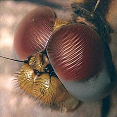
Compound Eyes (in insects and crustaceans) consist of up to several thousand light detectors called ommatidia, each of which has its own lens.
Single-lens eyes are found in vertebrates and some invertebrates.
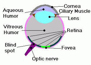 The eyeball in single-lens eyes is made up of two outer layers, the sclera (which in the front of the eye is the cornea-responsible for acting as a lens) and the choroid. The eyeball also contains the pupil, which is the hole in the center of the iris, and the retina, which contains the photoreceptor cells.
The sclera is an extension of the cornea.
Aqueous humor fills the anterior cavity of the eye, and the vitreous humor fills the posterior cavity of the eye.
The retina contains rod cells and cone cells, two types of photoreceptors.
Rhodopsin is the light-absorbing pigment that triggers a signal transduction pathway that ultimately leads to sight.
Identify the part of a single-lens eye with this link:
http://courses.washington.edu/chordate/453photos/skull_photos/human_ear_ossicles.jpg
Hearing and Equilibrium
There are three regions in the mammalian ear.
 1. The outer ear, which is the external pinna and auditory canal. These collect sounds and bring them to the tympanic membrane (eardrum), which separates the outer ear from the middle ear.
2. The middle ear, in which vibrations are conducted through three small bones collectively called ossicles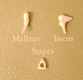 (individually, the malleus, incus, and stapes) and through the oval window.
3. Then the vibrations are conducted to the inner ear, which consists of a labyrinth of channels lined by membrane and containing fluid, all situated in bone.
The inner ear contains the cochlea, a two-chambered organ, which is involved in hearing.
The organ of Corti, which is in the cochlea, contains the receptors of the ear, which are hair cells with hairs that project into the cochlear duct.
The cochlea transduces the energy of the vibrating fluid into action potentials, in a wave that dissipates at the round window.
Some organs in the inner ear are responsible for detecting body position and balance. These are the semicircular canals.
~Quick Quiz~
1. The Ossicles in the ear contain three bones. What are they called?
a. the malleus, incus, and staple
b. the incas, mallus, and stapes
c. the malleus, incus, and stapes
d. the stepes, incas, and mallous
2. What part of the ear gives us balance?
a. semicircular canal
b. the pinna
c. the malleus
d. the incus
3. Which part of the eye reacts to color?
a. cornea
b. rod cell
c. rhodopsin
d. cone cell
answers: c,a,d
Mechanoreceptors are receptors stimulated by physical stimuli, such as pressure, touch, stretch, motion, or sound
Thermoreceptors respond to either heat or cold and help maintain body temperature by keeping the core temperature stable.
Chemoreceptors transmit information about solute concentration in a solution. Gustatory (taste) receptors and olfactory (smell) receptors are two types of chemoreceptors.
Photoreceptors and Vision
Compound Eyes (in insects and crustaceans) consist of up to several thousand light detectors called ommatidia, each of which has its own lens.
Single-lens eyes are found in vertebrates and some invertebrates.
The eyeball in single-lens eyes is made up of two outer layers, the sclera (which in the front of the eye is the cornea-responsible for acting as a lens) and the choroid. The eyeball also contains the pupil, which is the hole in the center of the iris, and the retina, which contains the photoreceptor cells.The sclera is an extension of the cornea.
Aqueous humor fills the anterior cavity of the eye, and the vitreous humor fills the posterior cavity of the eye.
The retina contains rod cells and cone cells, two types of photoreceptors.
Rhodopsin is the light-absorbing pigment that triggers a signal transduction pathway that ultimately leads to sight.
Identify the part of a single-lens eye with this link:
http://courses.washington.edu/chordate/453photos/skull_photos/human_ear_ossicles.jpg
Hearing and Equilibrium
There are three regions in the mammalian ear.
1. The outer ear, which is the external pinna and auditory canal. These collect sounds and bring them to the tympanic membrane (eardrum), which separates the outer ear from the middle ear.2. The middle ear, in which vibrations are conducted through three small bones collectively called ossicles
(individually, the malleus, incus, and stapes) and through the oval window.3. Then the vibrations are conducted to the inner ear, which consists of a labyrinth of channels lined by membrane and containing fluid, all situated in bone.
The inner ear contains the cochlea, a two-chambered organ, which is involved in hearing.
The organ of Corti, which is in the cochlea, contains the receptors of the ear, which are hair cells with hairs that project into the cochlear duct.
The cochlea transduces the energy of the vibrating fluid into action potentials, in a wave that dissipates at the round window.
Some organs in the inner ear are responsible for detecting body position and balance. These are the semicircular canals.
~Quick Quiz~
1. The Ossicles in the ear contain three bones. What are they called?
a. the malleus, incus, and staple
b. the incas, mallus, and stapes
c. the malleus, incus, and stapes
d. the stepes, incas, and mallous
2. What part of the ear gives us balance?
a. semicircular canal
b. the pinna
c. the malleus
d. the incus
3. Which part of the eye reacts to color?
a. cornea
b. rod cell
c. rhodopsin
d. cone cell
answers: c,a,d
Wednesday, April 22, 2009
Sensory and Motor Mechanisms
Hello children of AP Biology-

Today both blocks went over the most recent packet (chapters 46 through 48 I believe). Then, B, I think you guys started chapter 50. Ms. Lyon decided she was done teaching C block shortly after the packet, so we did nothing else. I'm assuming chapter 50 is one of our last... ah the beginning of the end. I'm just going to give everyone a quick overview of it. Also, chapter 50 vocabulary quiz is tomorrow as I'm sure you knew. Meghan kindly posted all the words yesterday, so I'll just pick several for a short quiz at the end. STUDY UP!
Chapter 50: Sensory and Motor Mechanisms
section 50.1
-basically, your body has two parts to its nervous system: the PNS and CNS
-the PNS is the peripheral nervous system (everything but the brain and spinal column): sensory reception occurs in this system
-a sensory receptor (made of specialized neurons or epithelial cells) detect stimuli--either internal or ext
ernal
-the receptor converts the stimulus energy to a change in t
he membrane potential of that receptor (look back at the action potential graph)
-this stimulus signal is passed along through action po
tentials to the CNS: the central nervous system (consisting of your spinal column and brain)
-the brain now goes through a process called PERCEPTION: it understands what is happening (the sound, smell, etc.)
-the signal is amplified and then the receptor cell either produces action potentials or releases a neurotransmitter
-there are numerous types of chemoreceptors (detect solute concentrations), mechanoreceptors (pressure, touch, stretch, motion, sound), electromagnetic receptors (electromagnetic radiation), thermoreceptors (temperature), and pain receptors
Alright guys, there's a baby intro to chapter 50. Here is a quiz over a few of the words for the vocab quiz tomorrow.
1. The broad region that corresponds to the length of the thick filaments of microfibrils is __________.
a) Z Line
b) A Band
c) I Band
2. The photoreceptor in the vertebrate eye that detects color during the day is _________.
a) Cone Cell
b) Trypomyosin
c) Rod Cell
3. A type of mechanoreceptor that detects sound waves and other forms of movement in air and water is _________.
a) Rhodopsin
b) Thin Filament
c) Hair Cell
Answers: (1) B (2) A (3) C
Good luck on the quiz!
shirts
Hey sorry I forgot to post this last night... but here are the potential bio shirts morgan and I designed. If you have any suggestions for changes, just post them as a response to this, and I'll get on it asap. I think all of C block is getting them. If anyone from B block wants one let me know.
-Linds
CLICK HERE to take a gander.
Tuesday, April 21, 2009
Nervous System
Hi ladies! Sorry for posting this so late. Don't forget that ch 50 is due tomorrow, the ch 46-48 take home is due tomorrow, and we have a ch 48/49 vocab quiz. IT'S ALSO LATE START! :)


Hydrostatic skeleton
a skeletal system composed of fluid held under pressure in a closed body compartment; the main skeleton of most cnidarians, flatworms, nematods, and annelids

rod cell
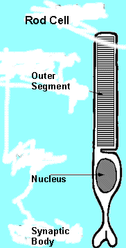
one of two kinds of photoreceptors in the vertebrate retina; sensitive to black and white and enables night vision


interoreceptor
a sensory receptor that detects stimuli within the body, such as blood pressure and body position

Roots:
 This is an action potential graph. An action potential or impulse can only be generated in the axon of a neuron. When the neruon is stimulated the sodium channels open and sodium ions flood into the cell. As a response, potassium channels open allowing potassium ions to leave the cell. This rapid movement (the upward line in the graph) is called depolarization. As you can tell, this lasts a very short amount of time. The potassium pump respores the membrane to its original polarized condition by pumping sodium and potassium ions back to their original location. This period is called repolarization. When the potential drops back down it goes through short period of time called the refractory period during which the neuron can't respond to another stimulus. Once the potential levels out, the neuron has reached it's resting state.
http://www.collegeboard.com/prod_downloads/ap/students/biology/ap04_frq_biology_b.pdf
Today in C block Lindsey showed us the awesome design for the AP Bio t-shirt. They are really cute and everyone should get one! She's going to post the link on the blog so that B block can see the shirts, too. We also took a vocab quiz and took more notes on the nervous system.
Chapter 50 Vocab:
Cardiac muscle
a type of muscle that forms the contractile wall of the heart. Its cells are joined by intercala ted disks that relay each heartbeat
Cardiac muscle
a type of muscle that forms the contractile wall of the heart. Its cells are joined by intercala
ted disks that relay each heartbeat
sarcoplasmic reticulum
a specialized endoplasmic reticulum that regulates the calcium concentration in the cytosol

a specialized endoplasmic reticulum that regulates the calcium concentration in the cytosol
Z lines
The borders of a sacromere
The borders of a sacromere
tropomyosin
the regulatory protein that blocks the myosin-binding sites on the actin molecules

the regulatory protein that blocks the myosin-binding sites on the actin molecules
A band
the broad region that corresponds to the length of the thick filaments of myofibrils

the broad region that corresponds to the length of the thick filaments of myofibrils
I band
The area near the edge of the sacromere where there are only thin filaments
The area near the edge of the sacromere where there are only thin filaments
sarcomere
the fundamental, repeating unit of striated muscle, deliminated
By the Z lines

the fundamental, repeating unit of striated muscle, deliminated
By the Z lines
Cone cell
one of two types of photoreceptors in the vertebrate eye; detects color during the day
one of two types of photoreceptors in the vertebrate eye; detects color during the day
thick filament
a filament composed of staggered arrays of myosin molecules; a component of myofibrils in muscle fibers
a filament composed of staggered arrays of myosin molecules; a component of myofibrils in muscle fibers
Hydrostatic skeleton
a skeletal system composed of fluid held under pressure in a closed body compartment; the main skeleton of most cnidarians, flatworms, nematods, and annelids
rod cell
one of two kinds of photoreceptors in the vertebrate retina; sensitive to black and white and enables night vision
thin filament
the smaller of the two myofilaments consisting of two strands of actin and two strands of regulatory protein coiled around one another

the smaller of the two myofilaments consisting of two strands of actin and two strands of regulatory protein coiled around one another
Myofilaments
the thick and thin filaments that form the myofibrils

Hair cell
a type of mechanoreceptor that detects sound waves and other forms of movement in air or water
a type of mechanoreceptor that detects sound waves and other forms of movement in air or water
rhodopsin
a visual pigment consisting of retinal and opsin. When rhodopsin absorbs light, the retinal changes shape and dissociates from the opsin, after which it is converted back to its original form

a visual pigment consisting of retinal and opsin. When rhodopsin absorbs light, the retinal changes shape and dissociates from the opsin, after which it is converted back to its original form
Amplification
the strengthening of stimulus energy that is otherwise too weak to be carried into the nervous system
the strengthening of stimulus energy that is otherwise too weak to be carried into the nervous system
pic n/a
Nocireceptor
A class of naked dendrites in the epidermis of the skin
Nocireceptor
A class of naked dendrites in the epidermis of the skin
interoreceptor
a sensory receptor that detects stimuli within the body, such as blood pressure and body position
Roots:
vitre-glass
tympan-a drum
sarco-flesh
sacc-a sack
omma-the eye
mechano-an instrument
coch- a snail
utric- a leather bag
tetan-rigid
rhodo-red
olfact-smell
ama-together
tropo-turn
noci-harm
stato-standing
gusta-taste
fovea- a pit
Graph from class:
This is an action potential graph. An action potential or impulse can only be generated in the axon of a neuron. When the neruon is stimulated the sodium channels open and sodium ions flood into the cell. As a response, potassium channels open allowing potassium ions to leave the cell. This rapid movement (the upward line in the graph) is called depolarization. As you can tell, this lasts a very short amount of time. The potassium pump respores the membrane to its original polarized condition by pumping sodium and potassium ions back to their original location. This period is called repolarization. When the potential drops back down it goes through short period of time called the refractory period during which the neuron can't respond to another stimulus. Once the potential levels out, the neuron has reached it's resting state.Questions from Ch 46-48 Take Home
1) Genetic mutations in asexually reproducing organisms lead to more evolutionary change than do genetic mutations in sexually reproducing ones because
A) asexually reproducing organisms, but not sexually reproducing organisms, pass mutations to their offspring
B) More genetic variation is present in organisms that reproduce asexually than is present in those that reproduce sexually
C) sexually reproducing organisms can produce more offspring in a given time than can sexually reproducing organisms
D) asexually reproducing organisms devote more time and energy to the process of reproduction than do sexually reproducing organisms
E) asexually reproducing organisms have more dominant genes than organisms that reproduce sexually
2) The Small portion of the embryo that will become its dorsal side develops from
A) morula
B) primitive streak
C) gray crescent
D) blastocoel
E) archenteron
3) Action potentials move along axons
A) by reversing the concentration gradients for sodium and potassium ions
B) more rapidly in myelinated than in non-myelinated axons
C) by the direct action of acetylcholine on the axonal membrane
D) by activating the sodium-potassium "pump" at each point along the axonal membrane
E) more slowly in axons of large than in small diameter
Answers: 1) A 2) C 3)A
Related FRQ
Related FRQ
http://www.collegeboard.com/prod_downloads/ap/students/biology/ap04_frq_biology_b.pdf
FRQ grading key
Beginning Ch. 48
An overview of the Nervous System
Sensory receptors collect information about the world outside the body as well as proscess information inside the body.
The Central Nervous System consists of the brain and the spinal cord. The conduction of signals from the central nervous system (CNS) to the effector cells is called motor output. Effector cells are target cells that produce some sort of response. They are generally gland or muscel cells.
The Peripheral Nervous System (PNS) consists of the nerves that commonicate motor and sensory signals in and out of the CNS. This reaches throughout the rest of the body. The PNS is divided into 2 groups
The Neuron is the functional unit of the nervous system. They are nerce cells. Neurons consist of 3 parts.
1. What are the basic building blocks of the brain?
A. brain cells
B. neurons
C. neurotransmitters
D. axons
2. What is most likely to be found in a synapse?
A. cells
B. neuron
C. neurotransmitters
3. Which is/ are true?
A. a neuron doesn't go through the cell cycle
B. a nephron transmits signals throughout the body
C. the 'fight or flight' response is atributed to the sympathetic nervous system
D. A & C
1. B ; 2. C ; 3. D
Sensory receptors collect information about the world
outside the body as well as proscess information inside the body.The Central Nervous System consists of the brain and the spinal cord. The
conduction of signals from the central nervous system (CNS) to the effector cells is called motor output. Effector cells are target cells that produce some sort of response. They are generally gland or muscel cells.The Peripheral Nervous System (PNS) consists of the nerves that commonicate motor and sensory signals in and out of the CNS. This reaches throughout the rest of the body. The PNS is divided into 2 groups
- Somatic nervous system- directs contraction of muscles
- Autonomic nervous system- controls organs and various involuntary muscles. broken into 2 parts
- Sympathetic nervous system- involved in stimulation of activities that ready the body for action (for example increasing heart rate)
- Parasympathetic nervous system- activates tranquil functions (stimulating secretion of saliva)
The Neuron is the functional unit of the nervous system. They are nerce cells. Neurons consist of 3 parts.
 Cell body- contaqins the nucleus and other organelles
- Dendrites- typically short, abundantly branched cell extensions that recieve incoming messages from other neurons
- Axon- a single , long, slim extension of the cell body that sends nerve impulses. Covering most axons is a myelin sheath which insulates and protects it. At the end of an axon is a synaptic terminal which transmits signals to other neurons via neurotransmitters, or chemical messengers
There are 3 groups that neurons are classified in:
- Sensory neurons- recieve initial stimulus Ex. those in the hand stimulated by touch
- Motor neurons- stimulate effector cells Ex. those stimulating sweat glands to cool the body
- Association neurons- located in the spinal cord or brain to recieve impulses from sensory neurons or send impulses to motor neurons. (aka interneurons)
The transmitting neuron is called the presynaptic cell and the recieving neuron is called the postsynaptic cell. Ganglia are clusters of nerve cells.
Reflex arc- a rapid involuntary response to a stimulus in which a sensory nerve  recieves information and passes it along to the spinal cord and then to a motor neuron, which signals an effector cell.
Glial cells are support cells. They are necessary for the proper functioning of nerve cells, and perform different functions depending on the type of glial cell.
- Astrocytes- provide support for neurons
- Oligodgendrocytes- form myelin sheaths around axons in the CNS
- Schwann Cells- form myelin sheaths around axons in the PNS
1. What are the basic building blocks of the brain?
A. brain cells
B. neurons
C. neurotransmitters
D. axons
2. What is most likely to be found in a synapse?
A. cells
B. neuron
C. neurotransmitters
3. Which is/ are true?
A. a neuron doesn't go through the cell cycle
B. a nephron transmits signals throughout the body
C. the 'fight or flight' response is atributed to the sympathetic nervous system
D. A & C
1. B ; 2. C ; 3. D
Subscribe to:
Posts (Atom)